What's new

Pořizování mikro klonů

newhousegreen

-.-
Veteran
Něco na přeložení::yoinks:
moc hezký:)

No, I have not done this nor do I know a lot about it. :)

I've been meaning to post this up for a while, it's collected info from the net and hope it helps anyone interested in tissue culture and artificial seeds.

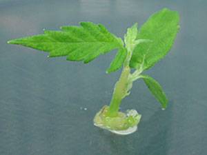
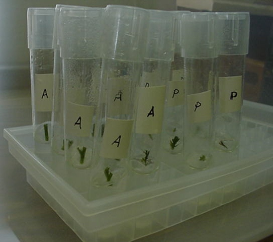
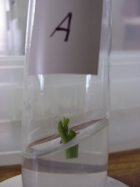
Micro cloning ie tissue culture is halfway to making the much coveted artificial seed. An artificial seed is basically a mini clone in a resin bead that can be stored, mailed etc. Although done with other plants, it has not been done with cannabis at a common level.

bioline_08pic1.jpg


bioline_08pic6.jpg


Chimera has done it. Thats the only person I know of.

artificial seeds would have some serious advantages - as far as storing parentals & mailing clones but their shelf life is currently limited, depending on encapsulation method - up to a year

Heres a link to what I am talking about: http://www.sp.edu.sg/schools/cls/bioline_08.htm

--------------

anyway, back to tissue culture - sterility is the most important factor

--------------heres a copy paste from another forum

Proper Auxin and Cytokinin concentrations need to be added to the mix during each stage.

Stage1--moderate levels of equaling auxin and cytokinin concentration--callus generation

Stage2--Transfer to media with incresed cytokinin concentration, and decreased auxin concentration to promote shoot formation.

Stage3--Transfer to media with incresed auxin concentration and decreased cytokinin concentration to promote root formation.

Stage4--Once roots are formed, the small plant is hardened off to lower and lower levels of humidity by letting the lid of the micro-propagation container open for longer and longer periods of time.

Stage5--Once the plant has hardened off to the dry atmosphere and can maintain osmotic balance, it is ready to transplant into soilless media. The amount of light must be monitored as well and a shade cloth may be needed for the first week.

Stage6--Once rooted in soilless media and after hardening off, the plant is ready for production. Small concentrations of fertilizer should be used initially, building up to the recommended rate.


**The most important thing in a successful tissue culture will be proper sterility of plant material, agar media, and the tools/person doing the transfers.
-
Ideally, the agar mix should be autoclaved for about 20-30 minutes to kill all pathogens. I have no idea if there is a way to autoclave in a conventional oven.... But if you do, be sure the glass container(s) with agar mix in them are not sealed tight because if they are they will likely burst with the increasing pressure inside from high temperatures.

Explants (plant tissue source) should be soaked in a 10% bleach-water solution for if I remember right about 2-3 minutes. The explant is then transfered into distilled water to rinse off.

All tools (tweezers) must be sterilized with ethanol (EtOH) and then quickly flamed at low heat to remove the ethanol.

Hands, arms, and everything on the working surface should be sprayed down with ethanol to prevent pathogen contamination in petri dishes.

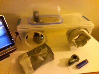
If you somehow have access to a HEPA filtered bench hood setup, this will increase the chances for success without pathogen growth on the media.....

============
more info:

This write up is directly about Cannabis
http://www.ib.uj.edu.pl/abc/pdf/47_2/145-151.pdf


http://www.growingedge.com/magazine/back_issues/view_article.php3?AID=190326

http://www.quisqualis.com/tv03tc01p1.html

http://web.telia.com/~u11206828/emetodik.htm


==========

some more info

Contaminating can be contained to a minimum with proper procedures, keeping things isolated. You'll never get 100% sterile, but you can get close enough that you can be very productive.

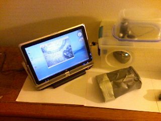
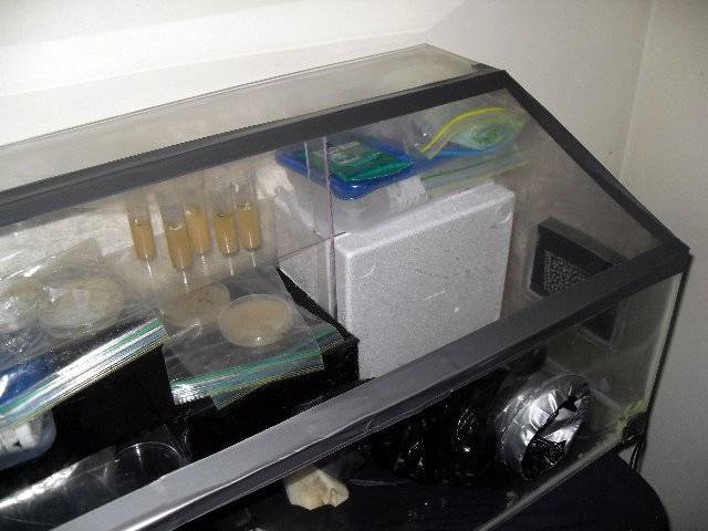
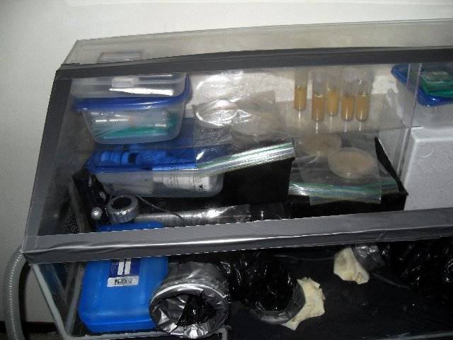
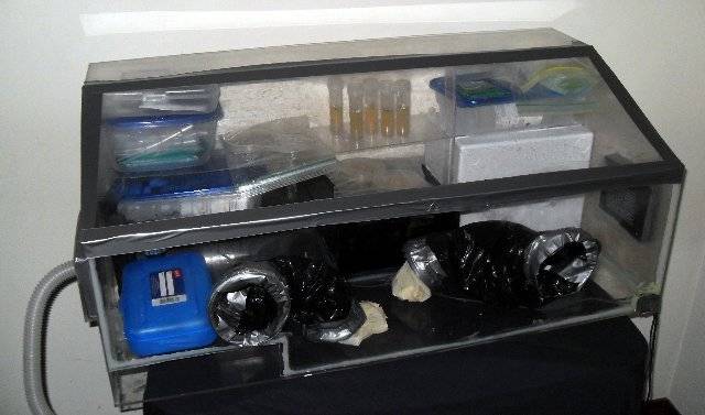
The more levels of containment the better. I do my cultures in petri dishes. The cells are under the agar and the agar has antibacterial additives, that's level 1... Level 2 is the dish itself. When you're not working with it, it should be sealed with gause tape. Perti dishes go in a new ziplock baggy, level 3. Level 4 is your clean box...

I only open the clean box to add or remove things, and I do it through a two-step process, so I can maintain relative cleanliness... If a glove breaks on me, I throw out every dish that isn't sealed.

here's some great books, but they r pricey, I think when I can, I will seriously get into this but that probably won't be for quite a while


51NRDEWDM9L._SS500_.jpg

http://www.amazon.com/Tissue-Cultur...=sr_1_1?ie=UTF8&s=books&qid=1228648531&sr=8-1


4111SZCAFCL._SS500_.jpg

http://www.amazon.com/gp/product/15...&pf_rd_t=101&pf_rd_p=463383351&pf_rd_i=507846


41cerYklmIL._SS500_.jpg

http://www.amazon.com/Plant-Propaga...=sr_1_4?ie=UTF8&s=books&qid=1228647736&sr=8-4


here's a video of someone who seems to know what they're doing

http://www.cannabistv.com/action/viewvideo/169/tissue_culture_media_prep/?ref=Loki777


387286940cannacallousregeneration.jpg


387284.png


387283.png


387282.png


387281.png


3872833.jpg


3872822.jpg


3872811.jpg


ok, now I have a question, in return. does anybody know if its true that tissue culture methods can restore the vigor to cuttings that are years old? or no? it's ok if nobody knows this, I'm just wondering.


.


Pokračování:http://icmag.com/ic/showthread.php?t=97494&highlight=agar
 

newhousegreen

-.-
Veteran
atd
This PDF guide was given to me by Carol Stiff (Ph.D)

Also can be down.oaded here http://www.zshare.net/download/17613900c740abeb/ :joint:


The PDF was 8 pages so I will do 8 post (if If the systems allows) to repersent each page.

TISSUE CULTURE AND AGROBACTERIUM-MEDIATED TRANSFORMATION OF HEMP
(CANNABIS SATIVA L.)
M. FEENEY AND Z. K. PUNJA*


Centre for Environmental Biology, Department of Biological Sciences, 8888 University Drive, Simon Fraser University, Burnaby,
British Columbia, Canada V5A 1S6
(Received 20 August 2002; accepted 30 April 2003; editor P. Ozias-Akins)


Summary​
Hemp (Cannabis sativa L.) is cultivated in many parts of the world for its fiber, oil, and seed. The development of new
hemp cultivars with improved traits could be facilitated through the application of biotechnological strategies. The purpose
of this study was to investigate the propagation of hemp in tissue culture and to establish a protocol for Agrobacteriummediated
transformation for foreign gene introduction. Stem and leaf segments from seedlings of four hemp varieties were
placed on Murashige and Skoog medium with Gamborg B5 vitamins (MB) supplemented with 5mM 2,4-
dichlorophenoxyacetic acid (2,4-D) and 1mM kinetin, 3% sucrose, and 8 g l21 agar. Large masses of callus were
produced within 4 wk for all cultivars. Suspension cultures were established in MB medium containing 2.5 mM 2,4-D. To
promote embryogenesis or organogenesis, explants, callus, and suspension cultures derived from a range of explant
sources and seedling ages were exposed to variations in the culture medium and changes to the culture environment.

None of the treatments tested were successful in promoting plantlet regeneration. Suspension cells were transformed with
Agrobacterium tumefaciens strain EHA101 carrying the binary vector pNOV3635 with a gene encoding phosphomannose
isomerase (PMI). Transformed callus was selected on medium containing 1–2% mannose. A chlorophenol red assay was
used to confirm that the PMI gene was expressed. Polymerase chain reaction and Southern hybridization detected the
presence of the PMI gene. Copy number in different lines ranged from one to four.​

Key words: callus; suspension culture; Agrobacterium tumefaciens; mannose selection; phosphomannose isomerase;
regeneration; transgenic hemp.

Introduction
Cannabis sativa L. is among the earliest cultivated plants and is
thought to have originated in Central Asia (Clarke, 1999). It is
valued as a food, oil, fiber, medicinal, and recreational drug source
and, consequently, has been dispersed throughout the world. Hemp
(Cannabis sativa L.) traditionally has been grown as a fiber crop and
there is a renewed interest in expanding its cultivation as a fiber and
seed crop in Canada. Hemp seeds possess high-quality oil and
protein (Johnson, 1999). Hemp varieties are now developed and
cultivated to produce high yields of fiber, seed, and oil, while
possessing negligible amounts of D9-tetrahydrocannabinol (THC),
the psychoactive compound, within the resin. However, the
confusion of hemp with marijuana varieties, which contain greater
amounts of THC, continues to hinder the widespread cultivation of
this crop (Forapani et al., 2001).
The development of new hemp cultivars with improved traits
could be further facilitated using biotechnological strategies. The
dioecious life cycle of many hemp varieties complicates breeding
efforts towards improvement of specific traits, such as resistance to
pests and diseases (Clarke, 1999).​


Development of a tissue culture system to regenerate hemp plantlets and an Agrobacteriummediated transformation protocol would permit exploitation of a
greater amount of genetic diversity for plant improvement and
would facilitate clonal multiplication of plants with desirable traits.
There are only a small number of reports concerning tissue
culture of hemp. Most of these studies were aimed at developing a
cell culture system to obtain secondary metabolites, particularly the
class of cannabinoids that are distinctive to the genus Cannabis
(Turner et al., 1980). Callus cultures (Hemphill et al., 1978;
Heitrich and Binder, 1982) and suspension cultures (Veliky and
Genest, 1972; Itokawa et al., 1977; Hartsel et al., 1983; Loh et al.,
1983; Braemer and Paris, 1987) have been established for
extraction of secondary metabolites and biotransformation studies.
Cryopreservation of hemp suspension cultures was developed as a
means to preserve germplasm collections (Jekkel et al., 1989). A few
reports have described tissue culture conditions intended for
plantlet regeneration. Richez-Dumanois et al. (1986) propagated
apical and axillary buds on stem explants in tissue culture and
subsequently rooted the shoots. A report by Fisse et al. (1981)
assessed organogenesis as a means of propagating hemp tissues.
They did not observe any direct organ formation on explants and
reported that Cannabis callus readily produced roots but was
unreceptive to shoot formation. Mandolino and Ranalli (1999) have
compiled an excellent review of the achievements with in vitro
*Author to whom correspondence should be addressed: Email punja@
sfu.ca
In Vitro Cell. Dev. Biol.—Plant 39:578–585, November–December 2003 DOI: 10.1079/IVP2003454
q 2003 Society for In Vitro Biology
1054-5476/03 $18.00+0.00
578​
atd
 

newhousegreen

-.-
Veteran
HEMP TISSUE CULTURE AND TRANSFORMATION​

hemp cultures with the objective of regenerating hemp plantlets.
There is only one account describing transformation studies with
C. sativa using Agrobacterium tumefaciens to transform shoot-tips
with a gene conferring resistance to infection by the fungal
pathogen, Botrytis cinerea (MacKinnon et al., 2000). However, to
our knowledge, there are no reports of somatic embryogenesis in
hemp.
The objective of this study was to identify the conditions for callus
and suspension culture growth of four fiber and seed hemp varieties,
to promote regeneration of plantlets via somatic embryogenesis
or organogenesis, and to develop an Agrobacterium-mediated
transformation protocol to introduce the selectable phosphomannose
isomerase (PMI) marker (Joersbo, 2001; Reed et al., 2001) into
hemp cells.


Materials and Methods

Plant material. Four hemp varieties representing different life cycles
and bred for either fiber or seed were chosen for this study. Varieties Uniko-
B and Kompolti are both dioecious and bred for fiber. Varieties Anka and
Felina-34 are monoecious and bred for seed and both fiber and seed,
respectively. Seeds were sown in 5 cm2 plastic containers containing
moistened potting mix soil (Sunshine Mix no. 1, Sun Gro Horticulture,
Bellevue, WA) at ambient room temperatures (21–248C). Seedlings
were placed under cool-white fluorescent lights with an intensity of
18mmolm22 s21 and a photoperiod of 12 h. Shoots were excised at the base
of the stem at 4 wk, at which time they had attained a height of about 20 cm
and had two to four pairs of true leaves.

The tissues were immersed in 70%
ethanol for 20 s followed by 10% commercial bleach (Javexw, containing
4.5% NaOCl) containing two drops of 0.1% Tween-20 per 100 ml for 1 min
while stirring. Tissues were then rinsed three times with sterile distilled
water and transferred to sterile Petri dishes lined with moistened filter paper.
Leaf (0.5 cm2) and stem segments (0.5 cm long) were excised after edges
were discarded and transferred to agar medium in 100 £ 15mm Petri dishes.
Callus induction. Leaf and stem explants of varieties Anka and Uniko
were placed on MB medium containing Murashige and Skoog macro- and
micro-nutrients (MS; Murashige and Skoog, 1962) with Gamborg B5
vitamins (Gamborg et al., 1968), 0.1 g l21 myo-inositol, 3% sucrose, 8 g l21
bacteriological agar (Anachemia Canada Inc., Montreal, PQ); the pH was
adjusted to 5.8 before autoclaving. A series of plant growth regulator
combinations were evaluated to induce callus development and somatic
embryo formation (mM): 2,4-dichlorophenoxyacetic acid (2,4-D; 2.5, 5, 9)
with either kinetin (0.5, 1, 5), a-naphthaleneacetic acid (NAA; 2.5, 5, 10),
6-benzylaminopurine (BA; 0.5, 1, 5), or indolebutyric acid (IBA; 2.5, 5, 10);
or IBA (2.5, 5, 10) with either BA (0.5, 1, 5), NAA (2.5, 5), or kinetin (0.5, 1,
5); or NAA (2.5, 5, 10) with either BA (0.5, 1, 5) or kinetin (0.5, 1, 5). Each
treatment consisted of 10 Petri dishes, containing 10 explants each. The
dishes were wrapped with Parafilmw and placed inside a dark drawer. After
1 mo, length and width of each callus mass was measured with a ruler and
averaged to obtain callus diameter.

Callusing responses of different explant sources were evaluated using
tissues from seedlings of varieties Felina and Uniko. The aerial structures
(leaves, petioles, stem, and cotyledons) from each seedling were sterilized
and sequentially cut into small segments as described above. The segments
were carefully arranged in sequential order in a Petri dish containing MB
with 5 mM 2,4-D and 1 mM kinetin (MB5D1K). Callusing was recorded over
a period of 2 wk.

The rate of callus development on stem and leaf explants of all four hemp
varieties on MB5D1K medium was recorded at 3–4 d intervals over a 1 mo.
period. Ten explants were placed in each Petri dish and there were 10 dishes
for each variety. At 4 wk, length and width of each callus mass were
measured with a ruler and averaged to obtain callus diameter. Callus was
maintained by transferring to fresh medium every 4 wk. The experiment was
repeated three times over a 2 yr period using the same batch of seeds.
Suspension cultures. Suspension cultures were initiated by cutting 4-wkold
callus masses into small pieces and transferring 0.5–1 mg of tissue to
150-ml Erlenmeyer flasks containing MB supplemented with 2.5 mM 2,4-D
and 3% sucrose; the pH was adjusted to 5.8 (MB2.5D). Cultures were shaken
at 115 rpm under ambient laboratory conditions and with 12 h d21 light at an
intensity of 10 mmolm22 s21. Every 2 wk, three-quarters of the spent
medium was replaced with fresh medium. By 4 wk, suspensions were
established and transferred to 250-ml Erlenmeyer flasks by suctioning about
1ml cell volume through a 3mm diameter pipette tip and transferring to
fresh medium. Suspension culture growth was measured at 3–4 d intervals
over a 1 mo. period and fresh weights of tissues were recorded after filtration.

Dry weight was obtained by incubating tissues at 408C, until constant mass
was achieved. The experiment was repeated three times over a 1 yr period.
Regeneration of plantlets. A range of treatments was evaluated to
determine whether hemp callus could be induced to regenerate plantlets
either through embryogenesis or organogenesis (Table 1).​


TABLE 1
SUMMARY OF TREATMENTS EVALUATED TO PROMOTE HEMP PLANTLET REGENERATION THROUGH EMBRYOGENESIS OR ORGANOGENESIS
Tissue sourcea Treatment
Callus, rhizogenic callus Subculture from MB plus combinationsb of 2,4-D, NAA, IBA, kinetin, and BA to growth regulator-free MB each month.
Callus Subculture from MB5D1K containing casein hydrolyzate (0, 100, 250, 500 mg l21), L-glutamine (3.4mM), or L-proline (15mM)
to growth regulator-free MB each month.
Callus, rhizogenic callus Subculture to half-strength MB with BA (0, 1, 5, 10 mM) and place in light (12 h d21) or total darkness for 2 mo.
Callus, rhizogenic callus Expose to MB plus BA (5mM) containing silver nitrate (0, 11.7, 47, 70.6mM) for 1 mo. in total darkness then to light for 1 mo.,
then subculture to growth regulator-free MB.
Tissue segments Expose to MB containing thidiazuron (0, 0.5, 2.5, 5, 1mM) for 4 d and then subculture to growth regulator-free MB.
Suspension cells Expose to half-strength MB liquid with thidiazuron (0.5mM) for 1 mo. and then subculture to growth regulator-free half-strength
MB liquid.
Suspension cells Expose to MB liquid with 2.5mM 2,4-D and 10210M salicylic acid for 1 mo. and then subculture to growth regulator-free MB
liquid.
Callus, rhizogenic callus Subculture to half-strength MS with 1% activated charcoal.
Callus Initiate and maintain on MB5D1K medium in the light (14mmolm22 s21) or in total darkness for 1 yr. Subculture to growth
regulator-free MB every 2 mo.
Callus Expose to MB5D1K medium and incubate at 48C for 2 mo. Subculture to growth regulator-free MB every 2 wk.
Tissue segments Expose to 1 M sucrose for 1–3 d and then subculture to growth regulator-free MB.
Suspension cells Expose to half-strength MB with 5% sucrose for 2 mo. and subculture to half-strength MB every 2 wk.
a Explants were taken from 7-d-old or 4-wk-old hemp seedlings from four hemp varieties. A range of explant sources (hypocotyl, epicotyl, cotyledons, petioles,
leaves, and immature flower buds) were used to initiate callus.
b Refer to Materials and Methods, callus induction section, for plant growth regulator combinations.​
...
 

newhousegreen

-.-
Veteran
Hemp Transformation
Bacterial strains and plasmids. Agrobacterium tumefaciens strain
EHA101 (Hood et al., 1986) harboring the binary vector pNOV3635 was
used for transformation. The plasmid pNOV3635 contains a coding region
for PMI under control of the ubiquitin promoter from Arabidopsis thaliana
(Ubq3) and the nopaline synthase terminator (NOS). The PMI gene for
selection of transgenic plants is between the left and right T-DNA borders,
and a spectinomycin gene resides outside of the borders for maintenance in
Escherichia coli and Agrobacterium vectors. Agrobacteria were inoculated
into 25 ml Luria-Bertani (LB) medium (Sambrook et al., 1989) supplemented
with 150 mg l21 spectinomycin and 50 mg l21 kanamycin. The suspension
was shaken at 288C for 2 d. Bacteria were harvested by centrifugation. The
resulting pellet was washed with hemp cell suspension medium (MB2.5D).
The pellet was then resuspended in 5ml MB2.5D containing 100mM
acetosyringone to a final OD600 nm 1.6–1.8. Agrobacterium was incubated in
the medium for 10 min prior to inoculating plant cells.
Transformation procedure. Anka suspension cells (1 ml packed cell
volume) were suctioned with a 3mm diameter pipette tip and transferred
with 4ml of MB2.5D into a sterile Petri dish. The hemp cell suspension was
inoculated with 5ml Agrobacterium suspension for 30 min. Hemp cells were
then collected on a 70mm diameter filter paper (No. 1; Whatman Int. Ltd.,
Cambridge, UK) by vacuum filtration. The filter was placed in a Petri dish
containing MB2.5D with 8 g l21 agar and incubated in the dark for 3 d at
ambient room temperature. Hemp cells were also incubated with
Agrobacterium lacking pNOV3635. Throughout the transformation experiment,
dishes were wrapped with Parafilmw. The transformation experiment
was repeated four times with one to six replicate dishes each.
Effect of mannose on callus growth. The concentration of mannose that
inhibited callus growth was assessed for the variety Anka to determine the
selection criteria for transformation experiments. A series of concentrations
of D-mannose (0, 1, 2, 3%) in MB5D1K, with and without sucrose, was tested.


Six callus pieces, each about 0.5 cm2, were aseptically placed in each Petri
dish, with three dishes per treatment. Dishes were wrapped with Parafilmw
and placed in the dark for 4 wk. Petri dishes were weighed at the beginning
and end of the 4-wk period to determine callus growth over a range of sugar
concentrations. The experiment was repeated three times. Data from one
representative experiment was subjected to a one-way ANOVA with means
separated using the Tukey–Kramer HSD statistical test (P ¼ 0.05).
Selection of transformants. After the 3 d cocultivation period, cells were
transferred to fresh filter paper and rinsed three times with a total volume of
200 ml MB2.5D. The filter containing cells was then transferred to MB2.5D
with 8 g l21 agar and 300 mg l21 Timentin (SmithKline Beecham, Oakville,
ON) and placed in the dark for 7 d to inhibit bacterial growth. Cells were
then selected by transferring small callus clumps (about 0.3 cm3) to MB2.5D
with 1% mannose, 300 mg l21 Timentin, and 8 g l21 agar. Dishes were
placed in the dark for 4 wk. Cell masses that continued to grow were
transferred to MB2.5D with 2% mannose and 150 mg l21 Timentin for 4 wk.
The transformation frequency (number of independent events obtained per
number of targets for which transformation was attempted) (Reed et al.,
2001) was determined for representative dishes of each experiment. Twenty
callus clumps (representing callus lines) were selected for replicates of each
transformation experiment and maintained on MB2.5D with 2% mannose
with subcultures made every 4 wk.

PMI assays. PMI assays were performed by placing 0.6 cm2 callus
masses into wells of a 24-well ELISA plate (Becton Dickinson and Co.,
Lincoln Park, NJ). Each well was filled with 600ml of assay medium consisting
of MB2.5D with either 1% mannose or 3% sucrose, 0.1 g l21 of the pH
indicator chlorophenol red (CPR, Sigma-Aldrich Chemical Co., Milwaukee,
WI), and 8 g l21 agar. The pH was adjusted to 6 prior to autoclaving, resulting
in a red-orange colored medium once dispensed into wells. Plates were
incubated in the dark at ambient room temperature for 3 d and color changes
in the wells were recorded. Between five and 13 callus lines from each of the
four transformation experiments were evaluated. Cells capable of metabolizing
the sugar source release acidic by-products into the medium, reducing the
pH and causing a visible color change from red to yellow (Kramer et al., 1993).
To determine if there was contamination of hemp callus with
Agrobacterium, all callus lines were incubated on LB medium at 288C for
1 wk and examined for bacterial growth.
Molecular analyses. Genomic DNA was extracted using a modified
protocol from Schluter and Punja (2002). Callus samples (100 mg) were
ground with 25 mg polyvinylpolypyrrolidone (PVPP), approximately 100 mg
sterile silica sand, 200ml DNeasy AP1 buffer, and 4ml RNase (Qiagen,
Valencia, CA) in a 1.5 ml microfuge tube with a plastic pellet pestle (Kontes
Glass Company, Vineland, NJ) attached to a hand-held drill, until a
homogeneous mixture was obtained. Another 200ml of buffer AP1 was
added to the mixture, vortexed, and DNA was isolated following the Qiagen
kit procedure. Primers used were described by Negrotto et al. (2000),
amplifying a product of approximately 550 bp in size. Primers consisted of
two 18-nucleotide sequences: PMI-1 50-ACAGCCACTCTCCATTCA-30 and
PMI-2 50-GTTTGCCATCACTTCCAG-30, and were purchased from the
Nucleic Acid-Protein Service Unit at the University of British Columbia
(Vancouver, BC). Each 25ml reaction for PCR contained 5ml of template
DNA, 50mM MgCl2, 20mM Tris, 50mM KCl, 200mM of each dNTP,
0.2mM of each primer, and two units of Taq polymerase (Invitrogen,
Burlington, ON). Amplification was carried out in a DNA Thermal Cycler
9700 (PE Applied Biosystems, Mississauga, ON). PCR conditions were those
chosen by Negrotto et al. (2000), with settings adjusted to 3 min at 958C
followed by 30 cycles of 30 s at 958C, 30 s at 558C and 45 s at 728C, with a
terminal elongation step of 5 min at 728C.

For Southern hybridization, hemp genomic DNA was digested with
HindIII (Gibco BRL Life Technologies, Burlington, ON) and electrophoresed
on a 0.8% agarose gel. DNA fragments were transferred to a nylon membrane
(Hybond-XL, Amersham Biosciences, Piscataway, NJ) by capillary transfer
with 0.4M NaOH (Koetsier et al., 1993). Hybridization was performed
according to the Amersham protocol for Hybond-XL membranes. The DNA
was hybridized to a 32P-labeled 550 bp PMI fragment obtained by PCR
amplification of plasmid DNA. Blots were exposed to X-ray film (Kodak
X-OMAT) at 2808C with an intensifying screen for 3–24 h.

Results and Discussion
Callus induction. Based on previous work (Loh et al., 1983;
Mandolino and Ranalli, 1999), a combination of MS salts with B5
vitamins (MB) was chosen in this study to promote callus and
suspension culture growth. However, Cannabis explants have been
found to respond favorably to both MS medium (Itokawa et al.,
1977; Fisse et al., 1981) or B5 medium (Heitrich and Binder,
1982; Braemer and Paris, 1987). Callus developed on leaf and
stem explants of hemp varieties Anka and Uniko for all treatments
containing 2,4-D within 4 wk after plating. Overall, treatments
containing 2,4-D supplemented with the cytokinins BA or kinetin
promoted the greatest callus growth and best appearance (data not
shown). Other treatments in which 2,4-D was replaced with IBA
or NAA as auxin sources induced an initial callusing stage
followed by development of a mass of rootlets covered in fine root
hairs after 4 wk (Fig. 1a). The promotion of rhizogenesis by NAA
was also noted by Fisse et al. (1981).

The combination of 5 mM
2,4-D and 1mM kinetin (MB5D1K) was chosen in this study to
promote prolific growth of pale yellow, friable callus (Fig. 1b);
FIG. 1. Hemp tissue culture and selection of transformed cells. a,
Rhizogenic callus after 1 mo. on MB medium supplemented with NAA or IBA,
instead of 2,4-D, as an auxin source. b, Callus growth on leaf explants on
MB5D1K medium. c, An established suspension culture of the hemp variety
Kompolti, showing cell aggregates. d–f, Callusing responses of different
explant sources on MB5D1K medium. d, Arrangement of stem and cotyledon
explants at day 0. e, Hypocotyl and epicotyl explants with comparable callus at 2 wk; cotyledons showed a poor ability to callus. f, Callus developing around
petioles and leaf midveins, followed by cut edges. g–i, Selection of Anka cells
transformed with pNOV3635 on MB2.5D with 300 mg l21 Timentin and 1%
mannose after 4 wk. g, Nontransformed cells are arrested in growth. h,
Transformed cells distinguished by their increased size compared to
untransformed cells. Dishes are 9 cm diameter (a–h). i, Transformed callus
on mannose medium, forming large, pale yellow callus protruding from small,
dark yellow parental callus (bar ¼ 5 mm).​
...
 

newhousegreen

-.-
Veteran
other studies have reported using a combination of 2,4-D and
kinetin for callus initiation (Hartsel et al., 1983; Braemer and
Paris, 1987) and establishment of suspension cultures (Braemer
and Paris, 1987).
Callusing responses on MB5D1K medium of different explant
sources from varieties Felina and Uniko were similar. However,
callus proliferation was dependent on the type of explant chosen,
and was greatest on stems, petioles, and leaves, and poor on
cotyledon explants (Fig. 1d–f). Fisse et al. (1981) and Mandolino
and Ranalli (1999) also noted that cotyledon and root explants did
not produce callus well. There did not appear to be any differences
in the extent of callus formation among leaves of different ages
taken from the same seedling (Fig. 1f).

The rate of callus development on MB5D1K for stem and leaf
explants of four hemp varieties was similar. By 2 wk, almost 100%
of the explants had produced callus and at 4 wk, the average callus
diameter was in the range of 6.8–7.8 mm, indicating that all four
hemp varieties responded similarly to MB5D1K medium (data not
shown). Callus from all varieties developed white roots covered with
fine root hairs if left longer than 4 wk without being transferred to
fresh medium.
Suspension cultures. Differences among the four hemp varieties
were more pronounced when tissues were transferred to liquid
MB2.5D medium. The extent of callus mass and root proliferation in
suspension culture differed with variety. Subcultures made every
2–4 wk favored development of small cell masses without rootlets
(Fig. 1c). Suspension cultures were easily established for the
varieties Anka, Kompolti, and Felina, while Uniko did not respond
well to suspension culture conditions. Suspension growth
experiments indicated that both fresh and dry weights of the
varieties Anka and Kompolti more than doubled within the first 7 d
of growth; however, Felina suspensions grew more slowly, and dry
and fresh weights doubled by 10–14 d, respectively (Fig. 2). Anka
suspensions produced the greatest fresh (8.50 g) and dry (0.63 g)
weights over 28 d.

Regeneration of plantlets. None of the attempts to regenerate
hemp plantlets, either directly from explants or indirectly from
callus or suspension cultures (Table 1), were successful. Neither
somatic embryos nor shoot bud initiation was observed in any
treatment tested. However, callus and suspension cultures had a
tendency to form roots. Fisse et al. (1981) and Hemphill et al.
(1978) described hemp callus which readily formed roots with no
evidence of shoot formation in response to different plant growth
regulator combinations. MacKinnon et al. (2000) also described root
development but did not observe shoot formation from hemp callus.
Alternatively, they developed a method to regenerate plantlets from
shoot-tips. Richez-Dumanois et al. (1986) developed a protocol to
micropropagate hemp using apical and axillary buds. With the
exception of Mandolino and Ranalli (1999), who described
occasional shoot regeneration from callus, de novo shoot formation
has not been reported for hemp. Reports of shoot or root formation
from callus as well as plantlet regeneration from shoot-tips or buds
suggest that hemp is capable of differentiation; further research
should be focused on this aspect to achieve an efficient and reliable
hemp regeneration protocol.

Hemp transformation. For our genetic transformation studies,
we used the PMI, manA, gene isolated from E. coli (Miles and Guest,
1984) as a selectable marker. The PMI selection strategy makes use
of a sugar (mannose) as a selection agent (Joersbo, 2001; Wright
et al., 2001) and is based on endowing transformed cells with a
metabolic advantage (Joersbo, 2001). Most explants are incapable of
growth on mannose as a carbon source (Joersbo, 2001) since this
sugar is converted by an endogenous hexokinase to nonutilizable
mannose-6-phosphate. In contrast, cells expressing the PMI gene
are capable of converting mannose-6-phosphate to fructose-6-
phosphate, which is readily metabolized (Negrotto et al., 2000;
Joersbo, 2001; Reed et al., 2001). This selection strategy has been
recently applied to several crops, including sugar beet (Joersbo et al.,
1998), cassava (Zhang et al., 2000), maize (Negrotto et al., 2000),
wheat (Wright et al., 2001), and rice (Lucca et al., 2001).
Medium supplemented with 1–3% mannose as a carbon source,
with or without sucrose, significantly arrested hemp callus growth
compared with medium containing 3% sucrose (Fig. 3a). Callus
placed on mannose had a similar appearance to callus growing on
sucrose, except that callus clumps were arrested in growth on the
former carbon source and were easily distinguished at 4 wk by
appearing much smaller and deeper yellow in color compared to
callus growing on sucrose (Fig. 1g–i). Medium containing
1% mannose therefore was chosen for selection of transformed
hemp cells.

ivyc0p.jpg


After Agrobacterium infection and cocultivation, callus developing
on MB2.5D with 1% mannose and 300 mg l21 Timentin turned
from pale yellow to a darker yellow color within 1 wk. By 4 wk, cells
capable of metabolizing mannose were easily distinguishable by
their color and larger size. Pale yellow callus emerged from darker
yellow cell clumps, growing larger than other callus masses
(Fig. 1h, i). These were transferred to fresh selection medium with
2% mannose and 150 mg l21 Timentin for proliferation. An average​
detected (data not shown). Two representative assays are shown in
Fig. 4. To evaluate control callus in the CPR assay, a 1 wk
pre-incubation period on mannose-containing medium instead of
sucrose was required. If control callus was grown on sucrosecontaining
medium, a change from red to yellow was observed (data
not shown). This suggests that growth on sucrose allows hemp cells
to store energy reserves that could be used for metabolic functions
that would acidify the assay medium, giving a false positive
reaction. Therefore, untransformed callus placed on MB2.5D with
sucrose as an energy source acidified the medium, turning CPR a
pale yellow color (lane AS), whereas untransformed callus
maintained on mannose-containing medium and transferred to the
PMI assay could not metabolize mannose and the assay medium
remained red (lane AM). Transgenic callus that grew on medium
containing sucrose or mannose (lanes T) changed the color to pale
yellow. Control wells without any callus remained red-orange
throughout the experiment. All callus masses absorbed a small
quantity of CPR dye from the assay medium, but this did not affect
subsequent callus growth.
Between five and 13 callus lines from each of four transformation
experiments were evaluated for PMI activity. All but two callus
lines turned the CPR medium yellow within 3 d. Presence of the
PMI gene was later confirmed by PCR analysis (see below).
In contrast, callus that had not been transformed did not induce a
color change over 3 d. Presence of the PMI gene within the two lines
not expressing PMI suggest that they may be low expressers and that
enzyme activity may not be detected. Wright et al. (2001) also
reported low-expressing transgenic tissue with mannose selection
and suggested that the PMI selection system can encourage growth
of both low- and high-expressing transgenic tissue.
All callus lines incubated on LB medium for 1 wk at 288C showed
no signs of bacterial contamination. Plates were kept for an
additional 1 mo. at room temperature without the appearance of
Agrobacterium.
Molecular analyses. PCR analysis was performed on 28 callus
lines representative of all transformation experiments, including all
callus lines from the CPR assay. All transformed lines tested by
PCR were shown to contain an amplified sequence of about 550 bp,
corresponding to the region between the PMI primers (Fig. 5).
Untransformed callus did not produce any bands.
Eight callus lines that had tested positive for the presence of the
PMI gene by PCR were analyzed by Southern hybridization. The
PMI gene was detected in transgenic callus and not in
untransformed control callus. A single HindIII restriction site is
present inside of the right T-DNA border in the PMI-containing

asi vše:):1help:
 

CrTecekk

Member
ono de o to ze nekteri botanici nazivaji rostliny soustavou bunek v kolonii ktere jsou bv simbioze tak jak sem nekde psal tak neni problem udelat z bunek rostlin explantat (pouze u nejakych rostlin se chova dane pletivo jako druh MO) a kultivovat je jako MO. nekde sem to aju videl lip v nejake bychli pokud najdu tak loadnu..jinak newhousegreen krute!!!! vidim ze te to zajima :) zaklad je digestor s vnejsim filtrem a nejaka ta zarivka:)
 

newhousegreen

-.-
Veteran
Mě by bohatě stačily ty klony,buňky moc malý...mikroskop,nářadíčko-by vyšlo draho
Hledal jsem na icmagu jen heslem Agar,takže se na netu udčitě skrývají i jiné´,možná i české informace
porozhlídnu se,a taky už bych se měl pustit do předběžnýho překladu:D
 

CrTecekk

Member
kdyz budes sikovny a rychli tak to muzes udelat bez digestore a mozna z 30 ti par nesplesnivy, za klad je mit ciste ruce vse vysterelizovane a byt rychlej..
 

newhousegreen

-.-
Veteran
mno,houby ozkoušej rychlost myslim dostatečně-jsou náchylnější že?...
už se na ně chystám dloouho,ale zatim nebyla motivace
 

CrTecekk

Member
snad ti to bjasni nejake otazky kdyz to projedes v poslednim odkazu je na 10. strane pekne schema ktere pletivo a co a jak....
 

Latest posts

Latest posts

Top